- 0 332 350 37 67
- bilgi@ezgiulu.com.tr
Laser Mole Treatment
Laser Mole Treatment
Moles and Mole Tracking
Skin cancer, medically known as "Malignant Melanoma", originates from pigment cells (cells that give color to the skin) and progresses very quickly. Malignant Melanoma is one of the most rapidly increasing cancer types in the world. While 30% of malignant melanoma lesions develop on a mole, 70% occur on a normal skin surface. It can be treated effectively when diagnosed at an early stage. However, if diagnosis is not made during this period, it quickly spreads to regional lymph nodes and then to the whole body, significantly eliminating the chance of treatment.
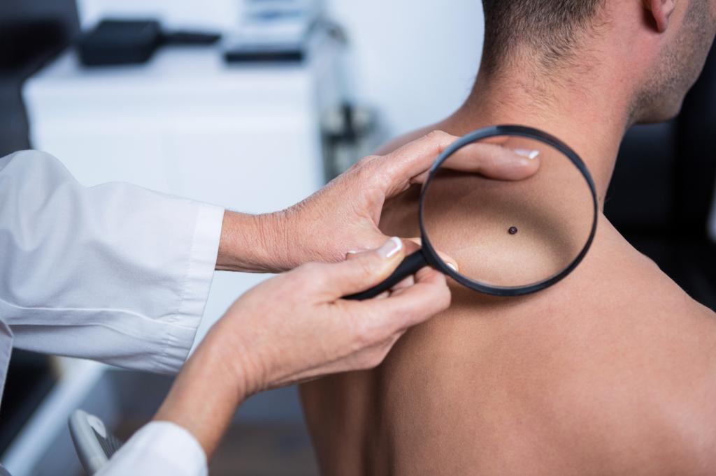
It is recommended that each individual examine their own moles monthly. Asymmetry, border irregularity, color change (mole containing more than one color), significant growth, inflammatory reaction or bleeding detected during this examination may indicate skin cancer. However, if a different-looking, fast-growing, pigmented lesion is detected, a dermatologist should be consulted immediately.
In the last 15 years, the risk of malignant melanoma has increased approximately 2-fold and, in parallel, new diagnostic methods have been developed. Dermatoscopy is skin surface microscopy; It is used in the diagnosis of moles and other pigmented lesions. In this method, the oiled skin surface is examined with a dermatoscope that provides light magnification. The dermatoscope is similar to the otoscope used to examine the ear and has been widely used for over 10 years. Until a few years ago, mole tracking was achieved by taking photos of dermatoscopic images. Then, digital dermatoscopy was developed by adding computer technology to this method.